
Behold: The First Medical X
Published: 7/14/2026 | Category: Search | Author/Source: Gizmodo.com
SpaceX’s Fram2 mission, launched in late March 2025, sent four amateur astronauts where no human had gone before. The flight will go down in history as the first to send a crew to polar orbit, but it will also be remembered for a major achievement in aerospace medicine.
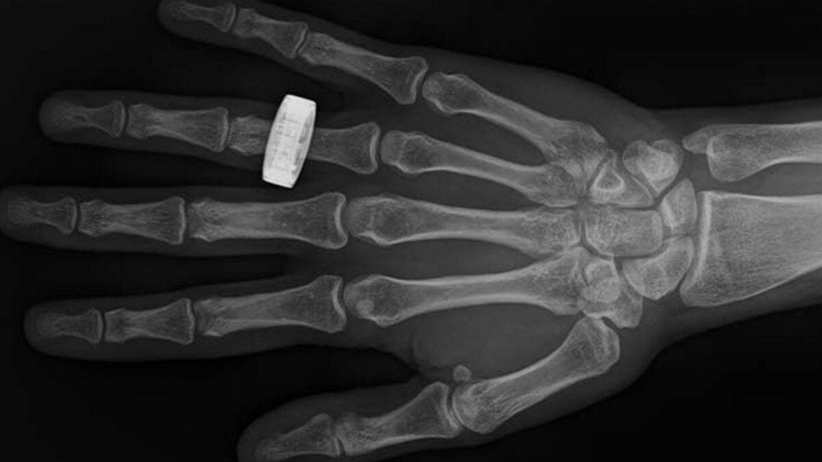
During their mission, the Fram2 astronauts took the first medical X-rays during an orbital flight. Without any guidance from ground control, they produced scans of a hand, forearm, abdomen, pelvis, and chest using a small, portable X-ray machine. The inflight images were immediately transmitted to an onboard computer to be reviewed by the crew, demonstrating that in-orbit radiography is feasible. The researchers unveiled them today in a study published in the journal Radiology.
For decades, ultrasound has been the only reliable medical imaging technique available to astronauts during spaceflight. But space is a dangerous place, and as missions grow longer and more distant, the risk of adverse medical events increases. Ultrasound, which requires considerable operator training and relies on a sound wave transmitting medium, may not always suffice.
“X-ray is one of the most powerful diagnostic tools in modern medicine because of its speed, accuracy, and ability to be operated by a broad range of people without the need of a sound transmitting medium,” lead researcher Sheyna Gifford, an assistant professor of aerospace medicine at Mayo Clinic, told Gizmodo.
“In the case of space, we would also be relieved to know whether or not our spacesuit glove had a hole in it, our rock pick was about to break from a stress fracture, and if the rock we picked up on our Moon walk contained the right minerals. A spectral X-ray system can help address all of these needs in the same set of equipment,” she said.
In 2022, Gifford co-authored a study that sent a portable X-ray machine on a parabolic flight, demonstrating that crew members could take viable diagnostic X-rays in a simulated microgravity environment. The next step was to test this capability in orbit.
Gifford’s team partnered with SpaceX to investigate whether astronauts could use a commercial off-the-shelf portable X-ray machine during the Fram2 mission, a 3.5-day polar orbital flight. Before liftoff, three of the crew members received four hours of operator training and acquired preflight images.
Fram2 launched aboard a SpaceX Falcon 9 rocket on March 31, 2025, putting a Crew Dragon capsule into a 90-degree orbit at 264 to 280 miles (425 to 450 kilometers) above sea level. During the flight, the crew used the portable X-ray machine to scan various body parts as well as a smartwatch, testing their ability to diagnose injuries as well as issues with electronics or equipment. The resolution of the smartwatch scan was down to the micron scale, Gifford said.
The mission returned on April 4, 2025, splashing down off the coast of Oceanside, California. The X-ray generator sustained some superficial damage during landing and recovery, but its internal hardware and X-ray output were unaffected. Once back on Earth, an operator who was not a member of the crew took postflight X-rays, replicating the preflight and inflight images.
Three independent radiologists evaluated all the images for quality, spatial resolution, contrast resolution, and positioning. Though their positioning differed somewhat, the images were the same across all other metrics, and the in-flight scans achieved a diagnostic level.
The study’s findings mark an important step toward expanding our diagnostic capabilities in space. Spacecraft such as the International Space Station (ISS) have long relied solely on ultrasound to assess astronaut health, but the imaging technique has limitations.
“Ultrasound, in the hands of a skilled technician, can detect some injuries and illnesses some of the time, with a variable amount of accuracy often after a good deal of looking,” Gifford explained. “For ultrasound to work, the injury or illness for which you are looking must be present in a medium responsive to sound waves.”
Because muscle, organ tissue, and arteries contain a lot of water, they are conductive to sound waves and show up nicely on an ultrasound screen. Bone appears, but with significantly less detail and clarity.
“A skilled ultrasound technician may know the best scanning angle to try for some small amount of sound wave penetration into bone, but the internal structure of the bone will often remain a mystery on ultrasound,” Gifford said.
The need to diagnose bone injuries in space will become even more important as humanity returns to the Moon.